
Endo Specialists
Recall Case
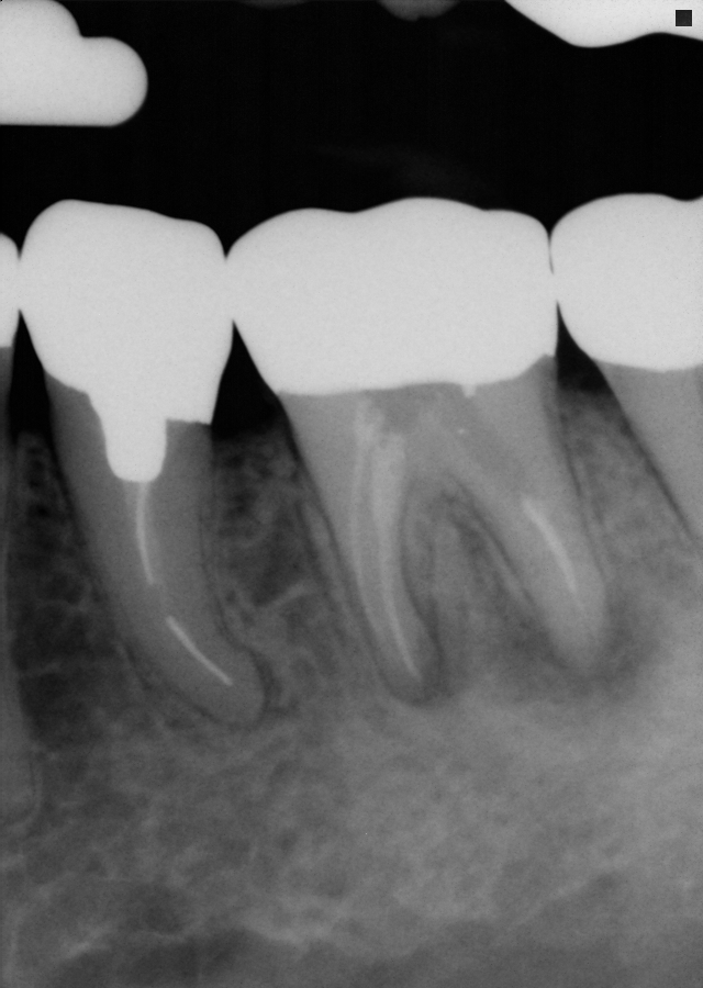
The following is a case of a 65-year-old male that was initially seen in January 2000. There was a RCT and a full gold crown on tooth #19. The patient has chewing sensitivity on tooth #19 and the PA reveals that there is a PARL on this tooth. The RCT obturation was short and thin on tooth #19. Tooth #20 has a crown with a silver point RCT. Tooth #20 was WNL to diagnostic testing and there is a widened PDL on this tooth.

Pre-op 01/25/2000
Tooth #19 was retreated using calcium hydroxide with was placed in the canals for two weeks.

Tooth #19 was obturated with gutta percha, Roth’s sealed and closed with cotton and Cavit.

Patient was seen for an evaluation of tooth #20 recently and this tooth will require an apicoectomy. Tooth #19 can also be seen on the PA and there is evidence of complete healing on tooth #19. Tooth #19 still has the same gold crown and it was sealed the access was sealed with an amalgam.
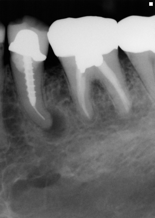
Pre-op Post-op
01/25/2000 09/24/2015
Resorption Case
The following is a case involving a 58 year old male patient that was asymptomatic on the lower right quadrant. The patient’s dentist referred him for an evaluation of tooth #30 which appeared to have a resorptive defect. A periapical image was taken of tooth #30.

Pre-op #30
Tooth #30 was positive to cold testing and WNL. Tooth #30 was negative to percussion and biting and the probing depths were WNL. The resorptive defect was clinically detectable on the buccal of #30. A CBCT was taken of the lower right area.

The diagnosis is invasive cervical resorption on tooth #30. The CBCT shows the extent of the resorption which can be seen in the coronal, sagittal and axial views. Based on the findings, it was determined that the resporption was external in nature but that it had extended into the pulp. Therefore, the RCT was initiated on #30 and four canals were cleaned and shaped. Calcium hydroxide was placed in the canals and the patient was to return in two weeks.

Intra-op #30
Tooth #30 was accessed, cleaned and shaped and the canals were obturated. After the canals were obturated, the resorptive defect was completely cleaned out with a #6 round bur. Glycerol was applied to the gingival tissues and then the defect was treated with 90% aqueous trichloracetic acid as described by Heithersay. The defect was curetted to remove the fibrovascular radicular extensions of the resorbing tissue.

Post-op #30
There were four canals present in tooth #30. Since the resorption did not cause a large external perforation, MTA was placed in the defect. The plan is to keep the existing crown and seal that access with a resin.